


INTRODUCTION
Vitamin D is known to play a part in a range of processes necessary for optimal health, such as the immune system, and the cardiovascular and musculoskeletal systems. Hence it is not surprising that many recent studies have examined the effects of its deficiency1.
Today, vitamin D deficiency is recognized as a widespread public health concern2. It has long been recognized that vitamin D contributes to the maintenance of both skeletal and non-skeletal health, as well as the balance of calcium and phosphate. Deficiency also plays a major role in metabolic bone abnormalities that result in adult osteomalacia and childhood rickets3. Furthermore, a lack of vitamin D has been associated with numerous chronic illnesses, including cancer, autoimmune disorders, heart disease and high blood pressure, conditions such as diabetes, metabolic syndrome, depression and neurocognitive function impairment, as well as heightened susceptibility to infection. As such, it is important to regularly test vitamin D blood level and follow up4-8.
Vitamin D is not categorized as a vital amine. It is a special pro-hormone that is primarily acquired through subcutaneous ultraviolet (UVB) light exposure. Briefly, 7-dehydrocholesterol (7-DHC) is converted to cholecalciferol (vitamin D3) in the skin after exposure to UV light. This is subsequently hydroxylated to the circulating metabolite 25-hydroxyvitamin D (25(OH)D). This form of vitamin D has a better absorption profile1.
Exposure to UVB is responsible for the creation of almost 90% of the vitamin D produced in the human body. Hence, lack of exposure to sunlight can result in vitamin D deficiency. The other 10% can be obtained from dietary sources such as cod, mushrooms, milk, eggs, and fortified foods. Although the production of vitamin D relies on exposure to UVB, the final level is also influenced by various other factors, including age, obesity, skin pigmentation, fashion choices regarding clothing, and the application of sunscreen9.
Recent research has also identified physical inactivity as a major risk factor for morbidity and mortality associated with chronic non-communicable diseases10, and for vitamin D deficiency11. A number of observational studies have found physical activity and exercise behaviour to correlate with vitamin D status12,13, and many indicate a significant positive correlation between physical activity levels and 25(OH)D levels14,15.
The core muscles are a collection of muscles that connect to the pelvis, hips and spine, which help support the spine and transfer force16. Neuromuscular trunk control can be improved in unstable situations by sling core stability training17; by creating training plans that adjust training intensity, increase trunk muscle strength and endurance, and activate the entire motion control system, it is possible to improve vitamin D absorption, together with stability and coordination18. Sling core stabilization training stresses closed-chain exercise under unstable conditions to improve movement control by the nervous system, strengthen the feedback between nerves and muscle groups, enhance the strength of deep stabilizing spinal muscles and improve balance; it is also believed to enhance vitamin D absorption19-20.
While vitamin D and calcium supplements have been well established to avoid or even reverse osteoporosis and sarcopenia in postmenopausal women, research indicates that load-bearing exercises like whole-body vibration (WBV) may offer the same benefits21-24. Indeed, WBV therapy has been found to impact the endocrine system, muscle function, bone metabolism and muscle training. It has been proposed that vibration therapy provides anabolic mechanical impulses to bone and muscle tissue, and that it increases the nutrient supply and vitamin D absorption by promoting blood circulation to the bones25. In WBV, the patient stands on an moving platform whose oscillations pass through the legs26; this movement stimulates subcutaneous proprioceptors, muscle spindles and Golgi tendon organs27,28. It has been proposed that WBV training may also improve vitamin D absorption29.
In Jordan, much of the population demonstrates low vitamin D levels, which have been linked to age, sex, obesity, and unemployment. There is therefore a pressing need for the local health authorities to raise awareness of vitamin D deficiency and how it can be prevented, especially among women30. Furthermore, a survey of Jordanian University students from non-health related faculties identified a need to improve the understanding, attitudes, and behaviors of the respondents related to vitamin D, its association with sunlight exposure, and the consumption of vitamin D-rich foods or supplements31.
Therefore, the aim of the present study was to compare the effectiveness of core stability exercise with that of whole-body vibration for improving vitamin D absorption in women.
MATERIAL AND METHODS
Study design
The study was performed at the Physical Therapy Department, Al-Zaytoonah University of Jordan. The study was authorized by the institution’s Research Ethics Committee under the number IRB#18\11\2024-2025. It was conducted between October 2024 and January 2025. Each participant received thorough information about the study’s objectives and methods, and provided their informed consent to take part, confirming their voluntary participation. The study was carried out in accordance with the 1964 Declaration of Helsinki and its later revisions. The study was also listed with the ClinicalTrials.gov registry with the registration number NCT06721637.
Participants
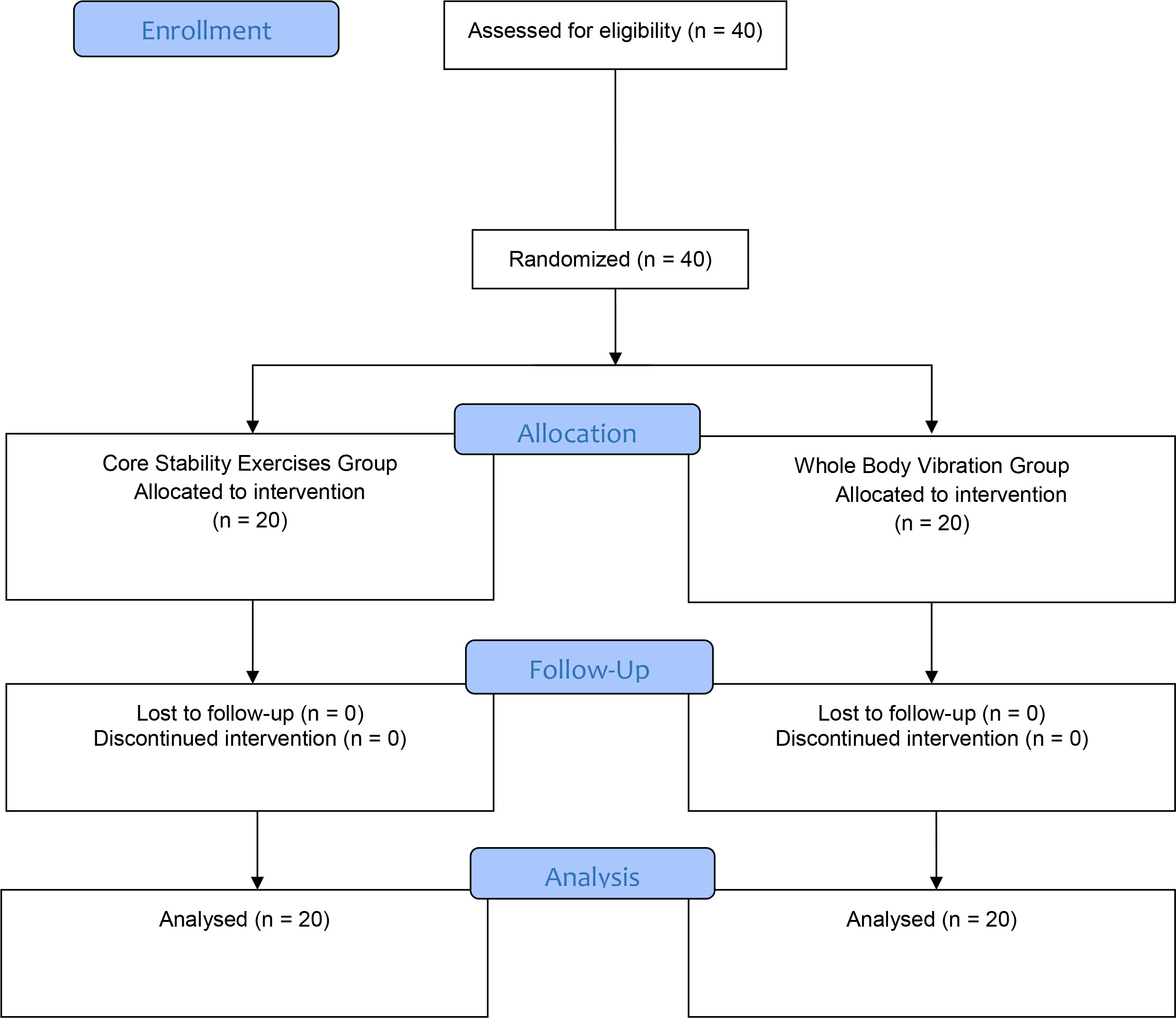
The study was carried out at Physical Therapy Department, Al-Zaytoonah University of Jordan. The inclusion criteria comprised the following: women, vitamin D deficiency, BMI <30, screening of vitamin D < 12 ng/ml. Participants demonstrating the following were excluded: a history of serious medical conditions like rheumatoid diseases, uncontrolled high blood pressure, severe heart or lung problems, or chronic viral infections like herpes and hepatitis, or who presented a vitamin D concentration higher than 12 ng/ml. All subjects in the study agreed to follow the same diet control and avoid any other types of exercise during the period of the study. The flow chart for the study is given in Figure 1.
Evaluation
Demographic data, weight, and height were recorded on a data sheet. The initial assessment of vitamin D concentration was performed based on parathyroid hormone (PTH) level. Vitamin D status is primarily assessed by measuring the level of 25-hydroxyvitamin D (25(OH) D) in the blood. This test, which involves a simple blood draw, provides a reliable measure of vitamin D level, and can be used to determine whether a person is vitamin D deficient, insufficient, or normal by comparison with reference ranges: a value below 12 ng/mL indicates deficiency, between 12 and 20 ng/mL insufficiency, between 20 and 50 ng/mL is generally considered normal. Normal PTH levels are generally between 10 and 65 pg/mL.
The weight and height of the participants were taken while they were wearing a thin layer of clothing. The BMI was then calculated by dividing the weight by the square of the height2 (kg/m2).
Intervention
The participants were randomly classified into two groups of equal size. Group A (20 women) received core stability exercise (CSE), and Group B (20 women) received whole-body vibration (WBV).
The intervention was performed three times a week for eight weeks, making a total of twenty-four sessions. All participants in both groups received a standard dose of vitamin D each day (880 IU/day); the first dose was given at the first physical therapy session and dosing was continued for four months.
Group A
Group (A) (n = 20): was given a vitamin D supplement in addition to the following core stability exercises; these were performed three times a week for eight weeks.
Abdominal hollowing exercise: the participant lay in a twisted posture, her feet resting flat on the treatment table. The therapist positioned two thumbs on the anterior superior iliac spines and directed the participant to contract her abdominal muscle for 10 seconds and then relax it for another 10 seconds32.
Quadruped abdominal hollowing exercise: the participant assumed a quadruped stance on the treatment plinth; the hips, knees and shoulders were flexed at 90 degrees and the spine was parallel to the table. The participant was instructed to breathe in and allow her belly to fall; as the participant breathed out, the participant drew her abdominal muscles toward her spine without moving her spine, maintained this for 10 seconds while breathing as normal. The participant then relaxed for ten seconds32.
Bilateral knee raise exercise: the participant assumed a lying position, resembling a crook. The participant was instructed to tighten her lower abdomen while maintaining normal breathing. The participant then raised her right leg toward her chest while keeping the contraction until it exceeded 90 degrees of hip flexion while allowing the knee to flex normally. The participant then held her right leg in this position and lifted her left leg in the same manner, so that both legs were elevated. The contraction was maintained for 10 seconds, then the right leg was returned to the starting position, followed by the left leg32.
Bilateral heel over exercise: the participant assumed the crooked lying position. The participant was instructed to contract her lower abdomen and to continue to breathe normally. While maintaining the contraction, she raised her right leg toward her chest until it just passed 90 degrees of hip flexion while allowing the knee to flex normally. While holding her right leg in this position, she then lifted her left leg in the same way, so that both legs were elevated. From this position, the participant was asked to extend both lower limbs toward the floor so that both heels were approximately three inches from the ground, without touching the floor with the feet. Both legs were extended until the knees became straight; during this move, the legs remained elevated approximately three inches from the treatment table. Then the knees were bent slowly towards the chest32.
The side bridge exercise: this exercise can be performed in one of two positions: upright against a wall, with the weight of the body supported on the hand, or on a table, with the body weight supported by a hand and knees and the hips elevated off the ground. This exercise has also been demonstrated to stimulate the lateral oblique muscles, which contribute to spine stability32.
Prone bridge exercise: when combined with the abdominal brace movement, this exercise activates all of the core musculatures. During the exercise, it is important to engage the gluteal muscles in order to maintain a neutral spine position. The participant was asked to lift her body by pushing on her elbows and tip of her toes32.
The exercises were performed at moderate intensity. The total duration of a typical core stability session was around 25-30 minutes, including the exercises and rest periods between sets. The patients were asked to start with shorter sessions and gradually increase the duration and intensity as they got stronger.
Group B
Group (B) (n = 20) received vitamin D supplementation and whole body vibration (WBV) training.
Fifteen minutes after the subjects arrived at the department, their blood pressure was measured and recorded. The participants were instructed to stand upright on the vibration platform during treatment, with both hands lightly gripping the handrails (Vibration Plate Trainer/ VIBRAFUN PRO / 150 kg/Taiwan). The body was maintained in an upright position with the focus shifted to the heel, ensuring that the vibration was conducted evenly upward; this was accomplished by stepping with a shoulder-width gait. The subjects then stood on the platform in various positions: half-squat, wide-stance squat, squat, deep squat, 1-legged squat, 1-legged stance, and lunge. At the conclusion of the vibration treatment, blood pressure was measured once more and the subjects were inquired about any discomfort. The treatment was given three times a week for ten minutes each session over the course of eight weeks. The linear vibration was performed at a frequency of 30 Hz and an amplitude of 5 mm33.
Statistical analysis
Intergroup comparisons of subject characteristics were made using an unpaired t-test. Normal distributions of data were checked using the Shapiro-Wilk test, and homogeneity of variance using Levene’s test. Mixed-design MANOVA was performed to compare within and between-group effects of training on vitamin D and parathyroid hormone. Subsequent multiple comparisons were performed using post hoc tests with the Bonferroni correction. The level of significance for all statistical tests was set at p < 0.05. All statistical analysis was conducted through the statistical package for social studies (SPSS) version 29 for windows (IBM SPSS, Chicago, IL, USA).
RESULTS
The characteristics of Group A and B are given in Table 1. No significant intergroup difference was observed in age, weight, height or BMI (p > 0.05).
Table 1
Basic characteristics of participants
The 2x2 mixed MANOVA identified a significant interaction between treatment and time (F = 27.7, p = 0.001, ƞ2 = 0.60). Significant main effects were observed for both time (F = 204.05, p = 0.001, ƞ2 = 0.91) and treatment (F = 26.5, p = 0.001, ƞ2 = 0.58).
A significant main effect was observed for treatment for Group A (F = 190.498, p = 0.001, ƞ2 = 0.911) and Group B (F = 41.311, p = 0.001, ƞ2 = 0.691).
Both groups demonstrated a significant decrease in parathyroid hormone and a significant increase in vitamin D between post-treatment and pre-treatment (p < 0.001) (Table 2). No significant difference was found between groups pre-treatment (p > 0.05). At the end of treatment, Group A demonstrated a significant decrease in parathyroid hormone and a significant increase in Vitamin D of compared with Group B (p < 0.001) (Table 2).
Table 2
Mean vitamin D and parathyroid hormone (PTH) values, pre and post-treatment, for Group A and Group B
The within-group analysis of vitamin D and PTH measurements is given in Table 3. Vitamin D was found to have a large effect size (0.947), with a Type III Sum of Squares value of 30,693.612. Parathyroid hormone also has a strong effect, with a Type III Sum of Squares value of 136,951.250 and an F-value of 941.595. In the between-group comparisons, vitamin D and PTH demonstrated lower Sum of Squares and significant F-values, indicating moderate effects. These results suggest significant differences in vitamin D and PTH levels, both within groups and between groups, with varying effect sizes.
DISCUSSION
The aim of the current study was to compare the absorption of vitamin D by women engaging in core stability exercise (CSE) with those receiving whole body vibration (WBV). Our findings indicate significant improvements in all measured parameters, including vitamin D and parathyroid hormone levels. The WBV and core stability exercises may have contributed to the notable increase in vitamin D levels observed in both groups. This is in line with Solymani and Habibian34 who propose that a combination of core stabilization and vitamin D intake can be effective in improving chronic low back pain in patients with low vitamin D levels.
Previous studies indicate positive correlations between bone density and the cross-sectional area of the lumbar abdominal core muscles; this is in line with the improvements in muscle development and BMD observed in both the intervention and control groups. Touban et al. report that long-term regular sling core stabilization training including calcium and vitamin D supplementation increased the cross-sectional area of the core muscles, improving BMD and vitamin D absorption35. Also, Ozsoy et al. found vitamin D treatment to enhance both balance and quality of life, while core strengthening and balance exercises also improved balance and prevented falls36.
Single workouts have also been found to raise blood vitamin D levels, suggesting that exercise may support vitamin D status. Regular exercise has been linked to increased expression of the muscle vitamin D receptor, which could improve the metabolism of vitamin D. This improvement is linked to better internal bone structure and mineral content, both of which are facilitated by increased vitamin D levels. vitamin D plays a key role in calcium absorption which is needed to maintain bone density37-38. Azeem et al.39 report that stability exercises appear to be of value in treating vitamin D insufficiency. Physical activity must be accompanied by adequate levels of vitamin D to increase bone density; vitamin D increases calcium metabolism by bones, which fortifies bones, reduces the risk of osteoporosis and enhances overall health. These studies highlight the value of combining regular stability exercises with sufficient dietary support, particularly vitamin D and calcium, in maintaining optimal bone health. This is particularly important in postmenopausal women. Such support can help women maintain their self-reliance, self-assurance, and social engagement, ultimately improving their general quality of life.
Our findings are in line with those of previous research on the effects of WBV on physical fitness in children and adolescents with disabilities . Their data indicates that WBV can elicit beneficial improvements in bone mineral density, muscle strength/power, gait speed, balance, lean body mass, and body fat40-42. In one study, Pin et al. review a number of prospective studies ranging from eight to 52 weeks in length41; it was suggested that WBV interventions lasting 10 to 20 min, performed three times a week, at higher frequencies (15–35 Hz) may be most effective for eliciting improvements in body composition and strength.
Other studies have investigated the effects of WBV training in conjunction with vitamin D supplementation on BMD and muscle strength. One randomized controlled trial examined the effects of six months of WBV training with either standard (880 IU) or high-dose (1600 IU) vitamin D supplementation among elderly women living in institutions found blood vitamin D levels, hip BMD and dynamic muscular strength to increase significantly in all groups. However, WBV did not demonstrate any appreciable benefits over vitamin D therapy alone. Moreover, boosting vitamin D did not improve musculoskeletal outcomes over the recommended dosage43.
Our findings support those of Li et al.44 who found bone density to be enhanced by the combination of WBV exercise and vitamin D administration. Additionally, vitamin D indirectly improved muscle strength, which contributes to overall body strength, making daily tasks easier and reducing the risk of falls. In addition, WBV exercise significantly raised vitamin D levels when taken as a supplement. Research indicates alarming levels of vitamin D deficiency worldwide, which has been confirmed by studies in Jordan15.
Our present findings confirm those of previous research indicating that vitamin D status is improved by a combination of physical activity and vitamin D supplementation. Both core stability exercises (CSE) and whole-body vibration (WBV) training were found to improve vitamin D absorption, and many previous studies have found it to improve bone health, muscle strength and balance in women.
While both CSE and WBV training are recommended to increase vitamin D absorption, they cannot be used as substitute therapy for pharmacological and dietary treatment methods. However, they may complement existing approaches.
Limitations of the study
The study involved only 40 participants, which may limit the statistical power and generalizability of the findings to broader populations. The eight-week duration may not capture long-term effects of exercise and supplementation on vitamin D metabolism or hormonal regulation. Both groups received exercise interventions alongside supplementation. Without a control group receiving only supplementation, it is difficult to isolate the specific impact of exercise.
CONCLUSIONS
While both the core stability exercise and whole-body vibration exercises were found to improve blood vitamin D and parathyroid hormone levels, better results were observed with the core stability exercise. However, further studies with follow-up examinations are needed to confirm these results.